
Osteoporosis
The skeleton acts as a rigid framework for the body by acting as a site for the attachment of muscles, protecting vital organs and by housing the bone marrow. Osteoporosis is a skeletal disease that is characterised by low bone density and deterioration of the bone tissue, which causes the bone to become more fragile and fracture much easier than normal. Regions of the skeleton can be replaced by bone remodelling, a process that involves the breakdown of old bone, which is reabsorbed by the body and then replaced by new bone. Under normal circumstances, the amount of old bone removed is balanced by the amount of new bone added. The main cause of osteoporosis is an imbalance in the remodelling process, where the amount of bone formed at certain sites is less than that which is reabsorbed or eroded.
Osteoporosis affects a large number of people 50 years and older, and is usually more common in women than men. The incidence of fracture rises steeply with age in both sexes, but occurs earlier in women than men. For example, the fracture rate in elderly women is twice that in men of the same age. Common osteoporotic fractures are of the spine and hip, but fractures of the forearm, wrist, humorous, tibia, ribs and pelvis also occur.
Symptoms
Osteoporosis may be characterised by pain, such as in the lower back, weight loss and abnormal curvature of the spine (kyphosis). New fractures may also give rise to severe pain, which typically decreases over several weeks to months. Pain is associated with localised tenderness and muscle spasm, which can limit movement. Individuals may also feel pain while standing up or during physical stress such as bending.
Diagnosis
Osteoporosis can only be diagnosed using a form of X-ray, called dual-energy X-ray absorptiometry (XDA) of the spine or hip, which measures the strength of the bone, or bone mineral density (BMD). However, other factors can also be used to predict the risk of future fracture, such as age, previous fracture and the use of certain medications can be used to identify individuals that may be most at risk.
Different types of fractures
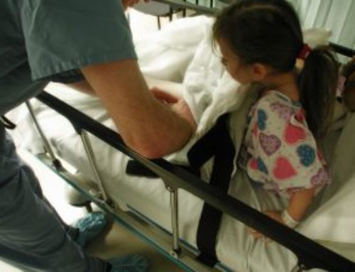
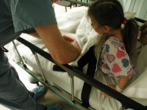
Hip fracture is the most devastating consequence of osteoporosis. For example, 5-20% of people die within the first year following hip fracture, and more than 50% of those sustaining hip fracture become incapacitated and require constant care. Hip fractures usually result from falling, but can also happen spontaneously. The risk of hip fracture also increases with age, which correlates with reduction in bone strength over time. For example, the majority of hip fractures occur in people who have fallen from a standing height and 90% occur in people who are over the age of 50. The incidence of osteoporotic hip fracture is lower in men than in women, which appears to be because men suffer fewer falls and are protected by having stronger bones, which lose less strength over time.
Vertebral fracture involves fractures of the spinal column, which can occur in 3 ways:-
- Crush (compression) fractures, where there is a loss in the posterior and anterior height of the spinal column
- Wedge (partial) fracture, where there is a loss in anterior height only
- Balloon (biconcave) fractures, where loss in bone tissue causes the vertebral end plates to become sunken or hollow
Vertebral fracture is reported to occur twice as often as hip fracture. They cause significant pain, deformity and long-term disability. Like hip fracture, the incidence of vertebral fracture rises with age, and is also thought to be much more common in women than men, although there is limited evidence to support this.
Distal forearm fracture involves fracture of the arm, which commonly occurs at the wrist following a fall onto the outstretched arm. This type of fracture is also associated with displacement of the radius and fracture of the ulna. Distal forearm fracture is also more common in woman than men.
Risk factors
The risk of an individual sustaining an osteoporotic fracture is determined in combination with the fragility of their bones and their risk of sustaining trauma, most commonly from a fall. Variation in BMD is the most common method used for assessing an individual’s future risk of fracture.
Bone mineral density (BMD) – is a strong predictor of future fracture. By measuring BMD at the site of interest the risk of osteoporotic fracture can be estimated. BMD is used in combination with other risk factors for osteoporosis.
Age and gender – are two of the strongest predictors of an individuals risk for fracture. As people age, their risk of fracture increases and compared to men, women are at greater risk. For example, 90% of hip fractures occur in patients above 50 years of age and of these individuals 80% are women.
Prior fracture – patients who have previously sustained one fracture are more at risk of subsequent fractures. For example, an individual who has sustained a prior vertebral fracture is nearly 4.5 times more likely to suffer from subsequent fractures.
Genetics – scientists have suspected a genetic component to osteoporosis and there appears to a consistent trend for heritability of some types of fractures such as in the lumbar spine and to a lesser extent, the radius and femur. Studies have shown that having a family history of fracture increases the risk of sustaining osteoporotic fracture. Genetic modelling has suggested that a number of genes contribute to the predisposition of individuals to osteoporosis and subsequent fracture. Numerous genes have been examined and those linked to osteoporosis include genes for the vitamin D receptor, estrogen receptor, growth hormones, collagen and insulin-like growth factor.
Biochemical markers – of bone formation and reabsorption can be used to measure the risk of fracture. Women increase the risk of developing osteoporosis following menopause, at which time, certain markers are known to increase, causing increased rates of bone loss. Individuals who have high rates of bone turnover can also be at increased risk of fracture.
Corticosteroids – increase the risk of osteoporosis by increasing bone loss. Rapid rates of bone loss and fracture occur in the first stages of therapy. Corticosteroids also reduce the capacity of the intestines to adsorb calcium and increase renal excretion of calcium.
Calcium – is one of the most important constituents of bone and the ability of the intestine to absorb dietary calcium decreases with age. This becomes most pronounced after the age of 70 and in women after menopause. Studies have shown that calcium supplementation increases BMD and reduces the risk of fracture.
Vitamin D – is involved in bone metabolism and is synthesised by the body following exposure to sunlight. As we age however, our ability to produce vitamin D from sunlight is reduced, and we become more dependent on dietary sources. Similar to calcium, supplementation with vitamin D increases BMD and reduces fracture risk. Good sources of vitamin D include fish such as salmon and tuna.
Other dietary factors – that tend to be associated with an increased risk of fracture include high caffeine intake, high protein intake, low vitamin intake and alcoholism.
Low body weight – is strongly associated with reduced BMD and increased risk of fracture. For example, individuals who suffer eating disorders such as anorexia nervosa are at increased risk of osteoporosis.
Smoking – tobacco reduces BMD by altering hormone metabolism, speeding up menopause, reducing calcium adsorption and reducing body weight.
Falls – landing on hard surfaces increases the risk of fracture, up to 90% of hip fractures occur from falling over. As people age, the risk of falling tends to increase the frequency of visual impairment or muscle weakness increases.
Prevention and management
The goal of prevention is to prevent the fractures that arise from osteoporosis. An important part of this process involves lifestyle advice to improve bone health. People can reduce their risk of developing osteoporosis and bone fracture by:-
- Exercising regularly. People with active lifestyles are at reduced risk of fracture. Slow lifting of heavy weights and high-impact exercise such as jogging or jumping are most effective at making bones stronger. Exercise also improves postural stability and helps to prevent falls.
- Adopting a healthy, balanced diet. High intakes of calcium, potassium, magnesium, vitamin D and vitamin K are important for maintaining strong bones.
- Reduce alcohol intake. Consuming more than 14 standard drinks per week can increase fracture risk. Modest alcohol intake (1-2 drinks per day) however, can actually have beneficial effects on BMD.
A growing number of treatments are now available for the management of osteoporosis. Nonpharmacolgic treatments include:-
- An optimal diet for the management of osteoporosis, including adequate calorie intake to avoid malnutrition, vitamin D and calcium. Excessive alcohol consumption should also be avoided.
- Quitting smoking to improve skeletal health
- Regular weight bearing exercise
- The use of hip protectors
- Orthopaedic management. Surgical treatment options can improve the health of people suffering from osteoporosis be increasing mobility and reducing pain.
Pharmacological treatments include:-
- Calcium and vitamin D supplement
- Hormone replacement therapy
- Synthetic estrogen analogs
- Bisphosphonates (reduces bone readsorption)
- Parathyroid hormone (increases bone formation)
- Strontium ranelate (increases bone formation)


Leave A Comment